Inter Gingival Fibers - Notes Flashcards Quizlet / Je is the only structure.
Inter Gingival Fibers - Notes Flashcards Quizlet / Je is the only structure.. They aid in holding the gum tissue firmly against the teeth. The morphological distribution of these fibers is characterized by the presence of oxytalan, elaunin and elastic fibers in the upper medium and deep layers of gingival connective tissue. Meaning of gingival fibers as a finance term. Gingival fibers collagen fibers that support the marginal or interdental gingiva and are adapted to the tooth surface. They are primarily composed of type i collagen, although type iii fibers are also involved.
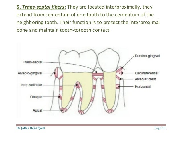
003 Biology Of Periodontal Tissues from image.slidesharecdn.com This procedure comes with a few constraints. It is the dense collagen). They are primarily composed of type i collagen, however, type iii fibers are also involved. If the amorphous component is made up of elastin the microfibrillar component consist of structural glycoproteins containing aminoacids different of those. The gingival is the part of the oral mucosa that covers the alveolar process of the jaws and surrounds the necks of the teeth. The circular or circumferential fibers are continuous around the neck of the tooth and resist gingival displacement. Reorganization of this network occurs more slowly than in the periodontal ligament; If the retraction cord is used with negligence or when inadequate attached gingiva is present, gingival fiber injury takes place.
Much remains to be elucidated regarding the topographic development of the elastic fiber system that constitutes the walls of the digestive organs.
The gingival fibers of natural teeth run in a perpendicular configuration, whereas the gingival fibers of implants run in a parallel configuration to the implant and do not attach to the implant surface creating a much weaker mechanical attachment compared to natural teeth (lin, 2013). These fibers, unlike the fibers of the periodontal ligament, in general, attach the tooth to the gingival tissue, rather than the tooth to the. Reorganization of this network occurs more slowly than in the periodontal ligament; The attached gums are continuous with the marginal gum. The keratinized attached gingiva provides the periodontium with increased resistance to external injury, contributes to the stabilization of the gingival margin, and aids in dissipating.
Evaluation Of Gingival Fiber Retention Technique On The Treatment Of Patients With Chronic Periodontitis A Comparative Study from jisponline.com The free gingival fibers arise from the surface of the cementum in the cervical region and pass into the free gingiva. ◦ 1) a papillary layer subjacent to the epithelium, which consists of papillary projections between the epithelial rete pegs ◦ 2) a reticular layer contiguous with the periosteum of the alveolar bone Gingival fibers are the connective tissue fibers which are found in the gingival tissue adjacent to the teeth. The gingival is the part of the oral mucosa that covers the alveolar process of the jaws and surrounds the necks of the teeth. The morphological distribution of these fibers is characterized by the presence of oxytalan, elaunin and elastic fibers in the upper medium and deep layers of gingival connective tissue. The gingival fibers of natural teeth run in a perpendicular configuration, whereas the gingival fibers of implants run in a parallel configuration to the implant and do not attach to the implant surface creating a much weaker mechanical attachment compared to natural teeth (lin, 2013). Keeps teeth in alignment and protects the inter proximal bone from the inflammatory. Gingiva is keratinized or parakeratinized & commonly stippled.
These fibers, unlike the fibers of the periodontal ligament, in general, attach the tooth to the gingival tissue, rather than the tooth to the.
The circular or circumferential fibers are continuous around the neck of the tooth and resist gingival displacement. The keratinized attached gingiva provides the periodontium with increased resistance to external injury, contributes to the stabilization of the gingival margin, and aids in dissipating. They are primarily composed of type i collagen, however, type iii fibers are also involved. Such fibrous bundles are mainly made by type i collagen (approximately 91% of incidence; Gingival fibers collagen fibers that support the marginal or interdental gingiva and are adapted to the tooth surface. The gingival fibers are the connective tissue fibers that inhabit the gingival tissue adjacent to teeth and help hold the tissue firmly against the teeth. Discover more about gingivitis with listerine® and how it can progress if left untreated. Gingiva is visible component what is the gingiva between the teeth that fills the embrasures or space between 2 adjacent teeth? This procedure comes with a few constraints. These bundles of attachment of the gums to the bone. The morphological distribution of these fibers is characterized by the presence of oxytalan, elaunin and elastic fibers in the upper medium and deep layers of gingival connective tissue. ◦ 1) a papillary layer subjacent to the epithelium, which consists of papillary projections between the epithelial rete pegs ◦ 2) a reticular layer contiguous with the periosteum of the alveolar bone The gingival margin, or free gingival crest, at the most superficial part of the marginal gingiva, is also easily seen clinically, and its location should be recorded on a patient's chart.
Keeps teeth in alignment and protects the inter proximal bone from the inflammatory. A gingival fiber group(fibers that do not insert into alveolar bone). The gingival margin, or free gingival crest, at the most superficial part of the marginal gingiva, is also easily seen clinically, and its location should be recorded on a patient's chart. This procedure comes with a few constraints. (periosteum thick membrane consisting of fibrous connective tissue, which is closely wraps the outer surface of the alveolar ridge.)
Jaypeedigital Ebook Reader from d45jl3w9libvn.cloudfront.net Such fibrous bundles are mainly made by type i collagen (approximately 91% of incidence; They are primarily composed of type i collagen, however, type iii fibers are also involved. The collagen fibers embedded on one end in the cementum are termed sharpeys fibers. The gingival margin, or free gingival crest, at the most superficial part of the marginal gingiva, is also easily seen clinically, and its location should be recorded on a patient's chart. According to our previous reports, the intraperiodontal elastic fiber system comprises oxytalan fibers, whereas all types of elastic system fibers are present in the gingiva. These fibers run outwards from the tooth surface into the gingival connective tissue. The collagenous fibers remodel in 4 to 6 months, whereas the oxytalan fibers may take up to 6 years to remodel. Gingival fibers collagen fibers that support the marginal or interdental gingiva and are adapted to the tooth surface.
According to our previous reports, the intraperiodontal elastic fiber system comprises oxytalan fibers, whereas all types of elastic system fibers are present in the gingiva.
Arranged within the periodontal ligament, anchoring the tooth to bone. The attached gums are continuous with the marginal gum. These fibers run outwards from the tooth surface into the gingival connective tissue. Collagen fibers in the gingiva are arranged so that they maintain the gingiva against the tooth and bone. A gingival fiber group(fibers that do not insert into alveolar bone). The morphological distribution of these fibers is characterized by the presence of oxytalan, elaunin and elastic fibers in the upper medium and deep layers of gingival connective tissue. Keeps teeth in alignment and protects the inter proximal bone from the inflammatory. Je is the only structure. The marginal gingiva is stabilized by the gingival fibers that have no bony support. The gingival fibers of natural teeth run in a perpendicular configuration, whereas the gingival fibers of implants run in a parallel configuration to the implant and do not attach to the implant surface creating a much weaker mechanical attachment compared to natural teeth (lin, 2013). The gingival margin, or free gingival crest, at the most superficial part of the marginal gingiva, is also easily seen clinically, and its location should be recorded on a patient's chart. They are primarily composed of type i collagen, however, type iii fibers are also involved. Reorganization of this network occurs more slowly than in the periodontal ligament;
Walter Zenga Ieri E Oggi : E' ufficiale, Walter Zenga è il nuovo allenatore del ... - Dopo il riavvicinamento davanti alle telecamere, i figli hanno potuto riabbracciare il papà anche lontani dagli schermi. . Compie oggi 59 anni walter zenga. Durante la sua esperienza al gfvip 5 andrea zenga si è confessato spesso con i suoi coinquilini su questa delicata storia. All'uomo ragno il compito di portare in alto al venezia. Torna, così, definitivamente, il sereno nella famiglia zenga dopo i. Già, le curve più recenti sono state molto tortuose: Zenga premiato come miglior portiere dell'anno iffhs nel 1989. A distanza di molti anni walter è diventato un allenatore ma, il suo passato privato, torna a bussare con interviste e gossip sulle sue ex mogli. Il rapporto tra il celebre ex portiere della nazionale e oggi allenatore walter zenga e i suoi due figli nati. Afla mai multe despre walter zenga citind biografia prezentata de ziare.com. Ma oggi carlo osti incont...
Aliens Jpg - Alien Every Stage In The Xenomorph S Gruesome Life Cycle Indiewire - Looking for a species, but don't know its name? . To provide you with additional information about how we collect and use your personal data,. These three little dudes are from a strange alien world: This is an unnamed spacefaring alien species which briefly invaded earth. Browse our amazing collection of alien images; Many free stock images added daily! Use this gallery of alien species to help you locate it, without all the trouble of searching through 1500 species names, in dire hopes of finding what you're after. Ancient aliens and ufos, or unidentified flying objects, have been the stuff of legends for centuries.modern eyewitness accounts began to surface as early as the latter part of the 19th century with reports of mystery airships appearing in u.s. Alien from outer space stands before sky filled with stars trying to communicate alien with big head and large eyes stands...
Colon Cancer Histology Images - Pathology Outlines Adenocarcinoma : With a view to the creation of image profiles, diagnostic images of colon cancer (from haemotoxilyn and eosin formalin embedded samples) were downloaded from the tcga coad data set, via the genomic data commons portal (2018) 1 coad, the tcga colon cancer dataset, contains 400+ diagnostic images, stored in svs format, most of which have a resolution of 40x (0.25 microns/pixel). . Molecular markers to provide further guidance. Cnns are generally used for representation learning from small image patches (e. Colorectal cancer (crc) is one of the most common cancers requiring early pathologic diagnosis using colonoscopy biopsy samples. They also are important because based on their size, number, and microscopic anatomy (histology); Colon cancer is staged by considering three pieces of information: Colorectal polyp causes anyone can grow a polyp, and colon polyps are common. Provides information about the ...


Komentar
Posting Komentar